Plug & Play: Plug in. Start endoscopy. Document.
In medical endoscopy, it’s not only the quality of image display that matters. Equally important is how easily a system can be integrated into everyday workflows. When examination, image transfer, and documentation are connected without unnecessary intermediate steps, individual components become a seamless solution.
This is exactly where orlvision’s plug-and-play principle comes in. The RSX series and the OX2 video otoscope feature USB connectivity and can be connected directly to suitable medical tablets or PCs. With the RSX series, digital image transfer is implemented directly via USB; with the OX2, image and video data are processed via a USB 2.0 interface.
USB instead of unnecessary system complexity
A classic endoscopy workstation can consist of several components. Camera unit, image processor, monitor, and other devices must be matched to one another. USB-based video endoscopes take a different approach.
The RSX series is designed for direct connection to a PC or laptop. With the RSX-USB, orlvision explicitly describes the USB connection as a plug & play connection to medical tablets and PCs—without a separate camera unit. With the RSX-HD, too, USB connectivity is part of the system integration.
This means: The endoscope does not first have to be integrated into the system via an additional camera unit. The image data can be transferred directly to the connected system and displayed or documented there.
This reduces the number of required intermediate steps and supports a clear, tidy workstation.
A USB video rhino-laryngoscope for immediate use
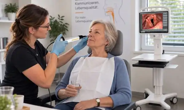
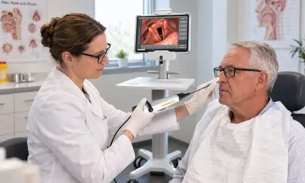
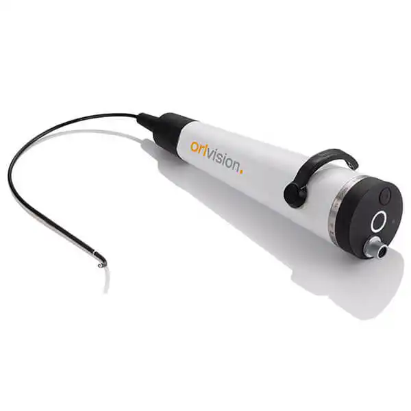
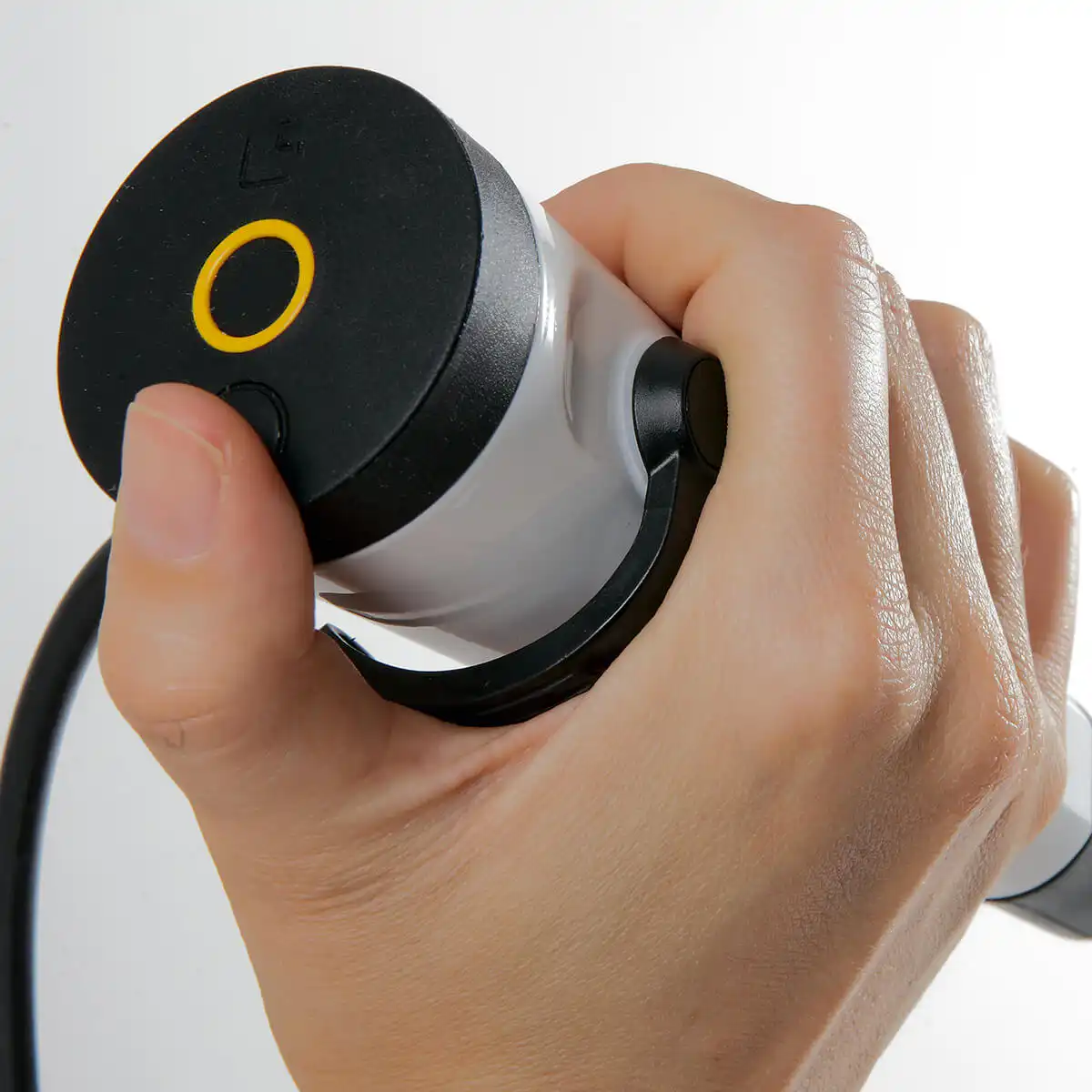
One example of this principle is the RSX-USB video rhino-laryngoscope. The compact video rhino-laryngoscope features a probe diameter of 3.9 mm, integrated LED illumination, and a direct USB connection. Digital image transfer enables immediate display of the examination and supports structured documentation of findings.
For the user, the focus is not on technical cabling but on the examination itself: connect, display the image, and document.
The other models in the RSX series also follow this concept. The RSX-HD combines a slim 3.9 mm insertion tube with a high-resolution image sensor and USB connectivity. With 800 × 800 pixels, it is designed for demanding diagnostic and documentation applications.
USB endoscopy for different requirements
For orlvision, plug & play does not mean that every endoscope is built identically. Rather, the USB interface connects different systems through the shared principle of direct digital connectivity.
For example, the RSX-P is designed with an especially slim probe diameter of 2.9 mm for pediatric and other sensitive applications. This video rhino-laryngoscope also features direct USB image transfer to medical tablets or PCs.
This allows the USB-based system concept to be combined with different examination requirements—from a compact standard system to a solution for particularly small anatomies.
USB not only for rhinoscopy
At orlvision, the plug-and-play principle is not limited to video rhino-laryngoscopy.
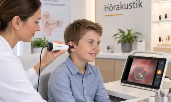
The OX2 video otoscope also features a USB interface. The system transfers the examination directly to a PC or monitor. The image and video data can be processed via USB 2.0 on a medical tablet.
The OX2 is another example of how a direct USB connection can simplify the technical integration of a medical imaging system.
From examination to documentation
A key element of digital endoscopy is the further processing of the captured image data. An examination does not necessarily end with the display of the live image. Images and videos can be relevant for documentation, reporting, or further communication within the medical environment.
That’s why the USB-based systems from orlvision should not be viewed in isolation. They can be part of a broader digital documentation environment. For example, the orlvision download page provides the orlView driver package with functions for image display and storage.
So the principle is not simply “USB instead of other connectors.” What matters is the connection between endoscope, image display, and documentation within a coordinated system.
Plug & Play for private practices, hospitals, and mobile applications
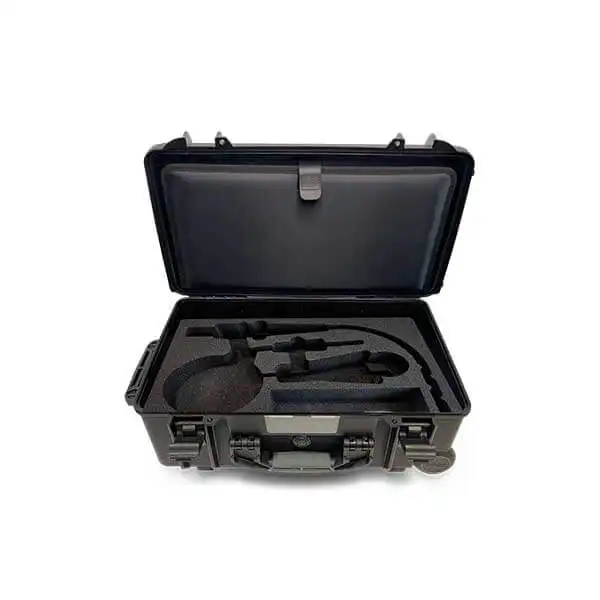
Another advantage of direct connectivity becomes apparent in mobile system concepts. The RSX series can be used together with a medical tablet in a transport case. orlvision describes this setup as a solution in which the tablet and endoscope can be transported together. This makes it possible to change the place of use without having to reassemble the entire setup.
This is particularly relevant when endoscopy is not intended to take place exclusively at a permanently installed workstation.
At the same time, the plug-and-play principle can also be integrated into stationary workstations. USB connectivity enables a direct connection to suitable medical PCs or tablets and can therefore be part of an individually configured endoscopy solution.
Individual products become a coordinated system
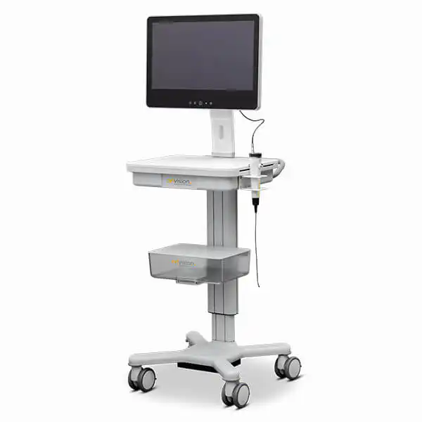
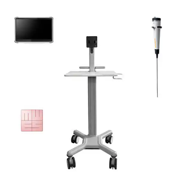
orlvision does not view endoscopy solely as a single device. The product page offers various endoscopes, tablets, holders, device trolleys, and other components that can be combined into individual systems.
The complete solutions on offer also demonstrate this concept. For example, a FEES system combines the RSX-HD with a medical tablet and a system trolley.
This turns a single USB endoscope into a solution that can be tailored to the specific workstation and the requirements of the medical application.
Plug in. Start. Diagnose.
This is exactly the core of the plug-and-play idea at orlvision: the technology should integrate into medical workflows without burdening users with unnecessary system complexity.
The RSX series combines video rhino-laryngoscopy with direct USB connectivity. The OX2 also transfers otoscopic images via USB. Different models allow adaptation to different examination requirements.
This turns simple plugging in into more than just a technical interface: USB becomes the connecting element between endoscope, image display, and digital documentation.
For medical professionals, this means a clearly structured workflow: connect the endoscope, display the image on the suitable system, and digitally process the examination further.
Sharp images. No detours.